在线客服1号


新品促销 快来看看吧
-
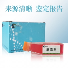
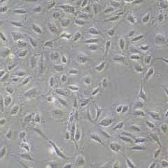
- 人皮肤成纤维细胞;BJ XY-H290
-
¥1700.00
-
-
-
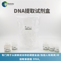
- 植物基因组DNA提取试剂盒 XY9701-50
-
¥550.00
- 植物基因组DNA提取试剂盒
-
- 快速转膜液(20X)
-
¥136.80
- 订单未满1000元,运费:20元
-
- 无血清细胞冻存液
-
¥228.00
-

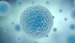
- 293T细胞 XY-H076
-
¥1100.00
- 人胚肾细胞
-
- BEAS-2B 人支气管上皮细胞 XY-H009
-
¥2125.00
- BEAS-2B细胞 人支气管上皮细胞 细胞